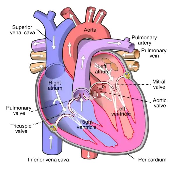
What is the name of the valve that separates the left atrium and left ventricle in the heart?
A. Aortic valve
B. Mitral valve
C. Tricuspid valve
D. Pulmonary valve
The mitral valve is located between the left atrium and left ventricle of the heart and helps to regulate the flow of blood between these chambers. It consists of two leaflets or flaps that open and close in response to changes in pressure as the heart beats.
During diastole, when the heart is relaxed and filling with blood, the mitral valve opens to allow blood to flow from the left atrium into the left ventricle. During systole, when the heart contracts to pump blood out of the left ventricle and into the systemic circulation, the mitral valve closes to prevent backflow of blood into the left atrium.
The mitral valve is one of four valves in the heart that help to ensure the unidirectional flow of blood through the heart and the rest of the circulatory system. Problems with the mitral valve, such as mitral valve prolapse or mitral stenosis, can lead to a range of symptoms and complications, including shortness of breath, fatigue, chest pain, and heart failure.
 |
Therefore, the Correct Answer is B.
More Questions on TEAS 7 Science
-
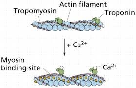
Q #1: What is the role of calcium in muscle contraction?
A. Calcium binds to tropomyosin to expose the myosin-binding sites on actin.
B. Calcium is released from the sarcoplasmic reticulum to initiate the sliding of actin and myosin filaments.
C. Calcium activates the motor neurons to stimulate muscle contraction.
D. Calcium is required for the relaxation of muscles after contraction.
Answer Explanation
Muscle contraction is a complex process that involves the interaction between actin and myosin filaments in the muscle fibers. The sliding of these filaments is initiated by the release of calcium ions from the sarcoplasmic reticulum, a specialized organelle in muscle cells. The calcium ions bind to the protein troponin, which causes a conformational change in the troponin-tropomyosin complex, exposing the myosin-binding sites on actin. This allows the myosin heads to bind to actin, forming cross-bridges that pull the actin filaments towards the center of the sarcomere, resulting in muscle contraction.
Option a) is incorrect because calcium does not bind to tropomyosin directly, but rather binds to the protein troponin, causing a conformational change in the troponin-tropomyosin complex. Option c) is incorrect because calcium does not activate motor neurons, but rather is released from the sarcoplasmic reticulum in response to an action potential that travels down the motor neuron to the neuromuscular junction. Option d) is incorrect because calcium is required for muscle contraction, not relaxation. The relaxation of muscles after contraction is due to the active transport of calcium ions back into the sarcoplasmic reticulum, which allows the troponin-tropomyosin complex to return to its resting conformation, blocking the myosin-binding sites on actin and ending the cross-bridge cycle.
-
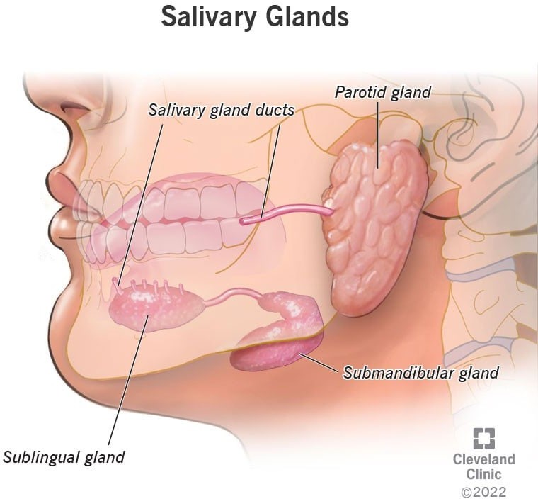
Q #2: What are the three types of salivary glands and where are they located in the mouth?
A. Parotid, sublingual, and submandibular glands located in the cheeks, tongue, and roof of the mouth, respectively.
B. Sublingual, submandibular, and buccal glands located in the tongue, cheeks, and lips, respectively.
C. Parotid, sublingual, and submandibular glands located in the roof of the mouth, cheeks, and under the jawbone, respectively.
D. Sublingual, parotid, and buccal glands located in the tongue, cheeks, and lips, respectively.
Answer Explanation
The three major pairs of salivary glands are the parotid glands, sublingual glands, and submandibular glands.
- Parotid glands are located just in front of your ears.
- Sublingual glands are located below either side of your tongue, under the floor of your mouth.
- Submandibular glands are located below your jaw.
-
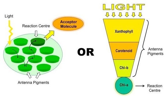
Q #3: What is the primary pigment responsible for photosynthesis in plants?
A. Chlorophyll a
B. Chlorophyll b
C. Carotenoids
D. Anthocyanins
Answer Explanation
Chlorophyll a is the primary pigment responsible for photosynthesis in plants. It is a green pigment that is essential for capturing light energy from the sun and converting it into chemical energy that can be used by the plant. Chlorophyll a absorbs light most efficiently in the blue and red parts of the spectrum, and reflects green light, giving plants their characteristic green color
Chlorophyll b is another type of chlorophyll that is also involved in photosynthesis, but it is not as abundant as chlorophyll a. Chlorophyll b absorbs light most efficiently in the blue and orange parts of the spectrum and reflects yellow-green light.
Carotenoids are pigments that are present in many plants and are involved in photosynthesis as well as protecting the plant from damage caused by excess light. Carotenoids are responsible for the orange, yellow, and red colors of many fruits and vegetables.
Anthocyanins are pigments that give plants their red, purple, and blue colors. While they are not directly involved in photosynthesis, they play a role in atracting pollinators and protecting the plant from damage caused by UV radiation.